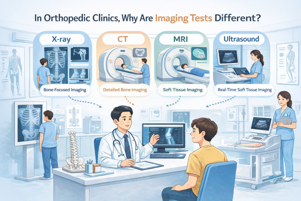
When you visit an orthopedic clinic, you may be advised to undergo different imaging tests such as X-ray, CT scan, MRI, or ultrasound, depending on your condition.
Many patients naturally ask:
“Why don’t we just do the best test from the beginning?”
“Isn’t MRI the most accurate one?”
These are very reasonable questions.
In this article, we’ll explain what each imaging test is best at detecting and why different types of scans are necessary in orthopedic care.
What Is an X-ray?
An X-ray uses a small amount of radiation that passes through the body to create an image based on tissue density.
- Bones, which block radiation well, appear white
- Soft tissues appear darker
What X-rays Are Best For
- Overall bone alignment
- Fractures
- Dislocations
- Joint space narrowing (arthritis)
Why X-rays Are Usually Done First
- Very quick examination
- Widely available
- Excellent for basic structural assessment
- Helps determine whether additional imaging is needed
In orthopedic practice, the X-ray is often the first step.
Just as an artist sketches the overall outline before adding details, orthopedic doctors first evaluate bone structure and alignment using X-rays before deciding on further tests.
How Is CT Different From X-ray?
A CT scan (Computed Tomography) uses multiple X-ray images taken from different angles, which are then processed by a computer to create cross-sectional (slice) images of the body.
This allows doctors to see bone structures in much greater detail.
When CT Is Especially Useful
- Complex fractures
- Small or subtle bone injuries
- Evaluating bone shape before surgery
Advantages of CT Over X-ray
- Three-dimensional understanding of bone anatomy
- Better detection of small or hidden fractures
CT is particularly helpful when precise bone detail is required.
What Is an MRI?
MRI (Magnetic Resonance Imaging) uses strong magnetic fields and radio waves to visualize internal tissues.
Importantly, MRI does not use radiation.
MRI is excellent for evaluating soft tissues.
What MRI Shows Best
- Cartilage
- Ligaments
- Tendons
- Muscles
- Intervertebral discs
- Other soft tissues
Key Features of MRI
- No radiation exposure
- Longer examination time
- Loud noise during scanning
- Very sensitive to body movement
MRI is often chosen when soft tissue injury is suspected.
Because MRI examinations take longer and are performed inside a narrow, enclosed tube, some patients with claustrophobia may find the test difficult.
Since the scan can be lengthy, remaining still for an extended period is also very important.
For patients with severe claustrophobia, or for elderly patients who may have dementia or delirium, the examination is sometimes performed with mild sedation to help them stay comfortable and still during the scan.
When Is Ultrasound Used?
Ultrasound uses high-frequency sound waves to create real-time images of internal structures.
It also involves no radiation exposure.
Advantages of Ultrasound
- Real-time dynamic evaluation
- Excellent for tendons, ligaments, and muscles
- Allows movement-based assessment
- Useful for image-guided injections, improving accuracy and safety
In orthopedic clinics, ultrasound functions almost like a stethoscope for musculoskeletal medicine.
Ultrasound is especially useful when performing injections.
In my own outpatient practice, I frequently use ultrasound-guided injections for patients with shoulder pain.
Using ultrasound allows accurate delivery of medication to specific structures such as the long head of the biceps tendon, the supraspinatus tendon, or the bursa.
Another advantage is that I can explain the findings to patients while showing them the ultrasound screen in real time, which helps them understand their condition more intuitively.
Common Questions Patients Often Ask
After learning about different imaging tests, many patients still have very practical concerns.
If you’re experiencing pain and are about to undergo imaging tests, you may find these questions helpful:
These articles explain common real-life situations patients often face in the clinic.
“Isn’t MRI the Best Test Since It’s the Most Expensive?”
Not necessarily.
Each imaging test has a different purpose.
- X-ray / CT → Bone-focused imaging
- MRI / Ultrasound → Soft tissue-focused imaging
Because they visualize different structures, no single test can evaluate everything.
The appropriate test depends on:
- Your symptoms
- Physical examination findings
- The suspected diagnosis
What’s More Important Than the Imaging Result?
Imaging Is Only One Part of the Diagnosis
- Some people show abnormalities on imaging but have no pain
- Others have significant pain even when imaging looks normal
Final diagnosis is based on:
- Patient symptoms
- Physical examination
- Imaging findings together
Images support clinical judgment — they do not replace it.
A Final Message From the Clinic
- “More expensive” does not mean “better”
- Imaging tests are not competitors — they each have different roles
- Choosing the right test step by step is usually the safest and most effective approach
Selecting the appropriate imaging study based on your current condition leads to better diagnosis and avoids unnecessary testing.
Leave a Reply